Richard Ernest Soldner was born in 1935 in Nurnberg, Germany. After graduating from school in 1950 he became trainee and toolmaker with Siemens AG between 1950 and 1954.
He received a scholarship from Siemens AG in 1955. From 1956-1960 Soldner completed a study of high-frequency engineering at the Ohm polytechnic institute, Nurnberg, the forerunner of the professional school. In 1960 he started work at the Department of Electromedicine at Siemens AG in Erlangen as development engineer in the area of accoustics, ultrasonic therapy and sonar metal flaw diagnostics. Other engineers who were there included Walter Erich Krause and Otto Heinz Kresse.
Siemens was a long-time maker of medical equipment, including X-ray apparatus. After Guttner had shown the impossibility of transmission imaging of the skull in 1952 (see Dussik), Siemens lost interest in diagnostic ultrasound. At around 1950 both Siemens and the smaller company Krautkramer, based in Cologne, started to make flaw-detecting equipment. Located close to the steel industry', Krautkramer had provided better service than Siemens, and ran a more profitable operation. Siemens decided to stop producing flaw-detection equipment in 1956. It was Carl Hellmuth Hertz who revived the company's interest in diagnostic ultrasound. Hertz adapted three of their flaw detectors in 1957 for cardiac investigations, and these were placed in hospitals in Germany. Sven Effert used it with good success in his hospital at Dusseldorf, and initiated ultrasound's use in echocardiography. Others such as S Blume contributed to some of the early electronics employed in the ultrasonic work.
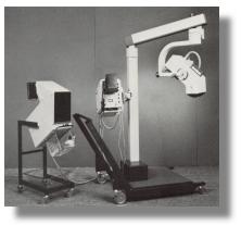
Directly into the making of B-mode scanners, Soldner and others at Siemens felt at that time the compound devices of Howry and Bliss in Denver and of Ian Donald in Scotland were too static to be diagnostically useful. They began work on an apparatus which will furnish a moving picture on the screen, which was particularly suited for Obstetrical work.
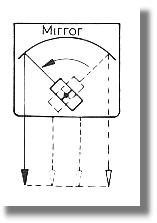
This led to the development of the device Vidoson, which had a relatively large, somewhat unmanageable ultrasound transducer head. By turning the transducer against a parabolic reflector, a wide useful picture in real-time sequences of more than 10 frames per second could be obtained. Initial testing was done at the University of Wurzburg and the image quality was however disappointing with both poor lateral resolution and penetration. The device took another two years to perfect. The final version had a much improved mirror and electronics, had 2 focused tranducers mounted on a small wheel and producing real-time images at 15 frames per second. The output was fed into an advanced Krautkramer oscilloscope.


Prototypes of these devices were field-tested by Hans J. Hollander, Gynaecologist at the University of Munster in 1964 and by E Gerhard Rettenmaier, Internist at the Erlangen Medical Clinic in the following year. The real-time device was received with great enthusiasm. The models 635/635S, 635ST, 735 and 735SM were commercially available from 1965 onwards. More than 3000 units were sold.
D Hofmann, Hans Holländer and P Weiser published it's first use in Obstetrics and Gynecology in 1966 in the German language. Hofmann and Holländer's paper in 1968 on "Intrauterine diagnosis of hydrops fetus universalis using ultrasound" also in German, is probably the first paper in the medical literature describing formally the diagnosis of a fetal malformation using ultrasound.
Malte Hinselmann, in Switzerland, using the Vidoson, demonstrated in 1969 the universal visualization of fetal cardiac action from 12 weeks onwards. The Vidoson was popular in the ensuing 10 years or so and were used in many scientific work published from centers in Germany, France, Switzerland, Austria, Belgium, Italy and some other European countries. The initial popularity was not based on its image resolution but rather its ability to allow the operator to display and study movements, such as fetal cardiac motion, gross body movements and fetal breathing movements. The device nevertheless did not sell well in non-German speaking countries and particularly in North America (see" Obstetric US imaging: the First 40 Years" by Dr. Barry Golberg).
Due to the handling problem of the large sound head with the Vidoson Soldner also developed in 1968 smaller handy linear array transducer heads. The group in 1971 put out improved versions of the scanner with multi-channel dynamic focusing using annula arrays. 1970-1977 further advancements took place in form of the device Echopan and also from doppler systems in form of the device Partecast.
Between 1977 and 1981 Soldner was active in the USA and participated in the system testing and special development of an ultrasonic transmission camera, which operated in the time of flight procedure. From 1981 up to his retirement in 1994, Soldner worked as a Senior engineer at Siemens AG, particularly in standardization procedures in IEC and TC 87. He also participated and in various ultrasonic reference committees in the federation KV in Cologne.
Today Soldner takes care of the devices of the ultrasound museum of the Deutsche Gesellschaft für Ultraschall in der Medizin (DEGUM) in Dresden and repairs the apparatus personally and keeps them in constantly functioning conditions. His efforts determined not only the developments at Siemens AG, but also the development of medical ultrasonics in general.
Read the Development of Ultrasound scanners at Siemens.
Read also a History of the development of ultrasonography, EFSUMB newsletters.
* Text and Images courtesy of and copyrighted DEGUMBack to History of Ultrasound in Obstetrics and Gynecology.