In a Presidential address to the Glasgow and West Scotland Obstetrical Society on 18th March, 1970, Donald said: 
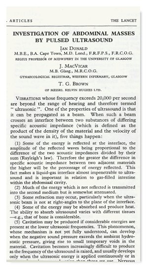
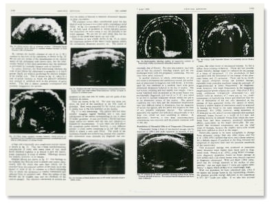
The front of the paper by Ian Donald, John McVicar and Tom Brown in the Lancet in 1958 (part of a 2-column page). It was considered as the most important paper on Obstetrical and Gynaecological sonography that was ever written. The paper spanned 9 pages describing the group's experience in 100 patients. It contained 12 illustrations of B-mode sonograms of the gravid uterus, ovarian cysts, fibroids and ascites and various normal and pathological conditions. It also depicted the prototype B-mode scanner gantry and probe and the Mark 4 flaw detector with which images were made. Safety of diagnostic ultrasound was also discussed in some detail.
" ..... At last we went to press and in 1958 the Lancet accepted an article which I wrote with John MacVicar and Tom Brown as co-authors and outlined not only the various principles upon which our work was based but showed a number of crude examples of our results. I think I shall always look back on that article as the most important I ever wrote and what is comforting is that all the conclusions which we reached at that time have since been vindicated by further results. The 200 reprints which I ordered were soon swallowed up and we had to resort to photostating to meet urgent requests ....... ."

Pages from the article showing B-mode images of the various pathologies.
An excerpt here of part of the Introduction and discussion will demonstrate the team's thinking and considerations at that time (1957-58). Although Donald's team had indeed been the world's pioneer in the design and use of the compound contact B-mode scanning device, and in particular its application in Obstetrical and Gynaecological patients, he had not claimed to be the first in the use of similar instrumentations and quoted works of Wild, Kikuchi and Howry :
" ......... A-scope PresentationBack to History of Ultrasound in Obstetrics and Gynecology.To confirm that echoes were obtainable within the body we started modestly with this method of presentation, which is standard practice in industry. By this method any echoes picked up are represented by vertical blips on a cathode-ray oscilloscope screen on a horizontal linear time-base sweep, the propagating source of ultrasound being represented by the left-hand end of the sweep. Since the velocity of propagation of ultrasound is very nearly the same in the various soft tissues encountered, the distance to the right along the base-line at which an echo blip is shown gives a measure of the distance of the reflecting interface from the propagating source…… The apparatus used was the standard' Mark 'IV' Kelvin Hughes flaw-detector, with which we had considerable experience, making 165 A-scope records of various solid and cystic swellings both in vivo and postoperatively in vitro.
It was not long before we discovered that the pattern of the blips is altered considerably by changing the angle of incidence of the ultrasonic beam, and it appeared that only the simplest reflecting interfaces could be diagnosed by conventional A-scope technique.
We were using at that time a standard frequency of 2 1/2 megacycles per second, and we compared the quality of echoes from the same subjects at 5/8, 1 1/4, and 5 mega-cycles as well, but we appeared to obtain the best results at 2 1/2 megacycles. The higher the frequency and therefore the shorter the wavelength the greater can be the resolution; but attenuation within the transmitting medium also increases with the frequency, as does "scatter", which confuses the picture, and therefore range is restricted for a given power. Reid and Wild (1952), using a higher frequency of 15 mega-cycles, were restricted to a range of about 2 cm only, but they suggested that this could be extended by the principle of time-varied sensitivity, in which the receiver gain is increased as a function of the elapsed time between propagating signal and echo. A compromise has to be reached between frequency and resolution and depth of penetration.
The use of A-scope presentation has been applied in ingenious ways, especially by Effert et al. (1957), who studied the movements of the left atrial walls of the heart simultaneously with electrocardiography and phono- cardiography in the relative assessment of mitral stenosis and incompetence. They also demonstrated pericardial effusion.
........... B-scope PresentationIn this arrangement the direction of the probe is kept constant, and the area is scanned by moving the probe sideways along a line at right angles to the ultrasonic beam. The display on the cathode-ray tube is made to follow this sideways movement of the probe. If the size of the echo is represented by variations in brightness of a spot on the cathode-ray screen instead of as a blip, it is possible to build up a composite picture on a long- persistence screen or on a photographic plate. A variant of this method was used by Wild and Reid (1951) using a hand-held instrument in which a propagating crystal scanned to and fro over a range of 6.5 cm. within an elliptical water-chamber. Another variant is to place the test object in a water-tank round the outside of which a probe travels directing a radial beam inwards. Similarly, in Japan, Kikuchi et al. (1957) graduated from A-scope presentation to a type of B-scope scan, which they call "ultrasono-tomography", for the exploration not only of the abdomen but also of the cranial cavity, as much of the head as possible being placed in a water-tank. The method appears to be still under development. Our experience was like that of Howry (1955), who m noted that even quite small angular displacements from the perpendicular in the incidence of the ultrasonic beam produced very great differences in the amplitude of the reflected echo. Howry calculated that an angle as small as 6° off the perpendicular reduced the amplitude of the detected echo to a tenth, and 12° reduced it to a hundredth. Howry and his colleagues have done a lot of fundamental work with simple geometrical test objects in water-tanks, and have used ultrasonic lenses to narrow the beam and to improve penetration and resolution. They have also attempted three-dimensional and stereoscopic observations of body structure by building up two composite pictures us taken from angles differing by 10° in respect of each other and studying them in a stereoviewer (Howry et al. 1956).
....... Radial Scan or Plan-position IndicatorA plan-position-indicator (P.P.I.) display is produced lat by rotating the probe about a fixed point in or near the ed area being scanned and causing the time base sweep to follow the angular movement of the probe while originating from a fixed point on or off the face of the cathode-ray tube. If the origin of the display is off the face of the tube, the display is known as a "sector scan". None of the Its aforementioned methods is sufficient by itself to produce a satisfactory echo pattern of a deep-seated structure within the body, because echoes are only detected by the receiving crystal if the reflecting surfaces from which they originate are at right angles to the incident energy beam. For these reasons it was decided to attempt to produce a scanning and plotting mechanism which, so far as of possible, would enable each individual part of the surface be of the structure under investigation to be scanned by the ultrasonic beam from a large number of different angles, all, however, lying in the plane of the cross-section to be represented. In this way we have sought to reproduce a composite cross-sectional view of the parts of the body c examined, "collecting" echoes on the one picture from as many angles as possible, registering simultaneously not only the echoes and their strength but also the position of the probe and the angle of the incident beam. Our apparatus thus combines B-scope and P.P.I. presentation.
.......... Discussion......... To be of any use at all to the clinician, the echo patterns obtained by pulsed ultrasound must be not only intelligible but also consistently reproducible at the same ti level in the same case. That this is not always so we attribute to inadequate scanning methods and technique -- hence the development of our combined B-scope and P.P.I. presentation and scanning. Even so we are very far from satisfied with the crude results so far obtained. Other workers have made claims which, in many instances, are more striking than substantial, judged by some of the illustrations offered. The temptation to try to distinguish benign from malignant tissue by such simple, quick, and harmless means is well-night irresistible; but, being only too well aware of the difficulties which even the histologists on direct microscopy may have in assessing malignancy, we have not attempted this sort of assessment in y the present state of crudity of an ultrasonic beam whose d width is measurable in millimetres and whose wave-length is many times the diameter of any living cell, benign or malignant. Wild and Reid (1952) claim that is it is possible to distinguish between a benign and a malignant tumour within the breast on the basis of positive n differences observable on ultrasonic echography. The Japanese workers Kikuchi et al. (1957) have made the same claim; they also claim to have recorded echoes from human intracranial ventricles, as we have, in the newborn, and they make other claims which we have not explored. Howry and Bliss (1952) noted that fresh specimens in vitro differed in their sonic properties when compared with fixed specimens in formalin solutions. We also have noted differences between the tumour in to vivo in a tank after removal from its host. We cannot explain this difference except by suggesting that blood lrs circulating through a tumour alters its sonic properties.
Our experience of 78 cases in which diagnosis was he quickly verified by laparotomy and subsequent histology he indicates that ultrasonic diagnosis is still very crude, and that the preoperative diagnosis of histological structure is still far off, although such a possibility in the future is an exciting prospect. The fact that recordable echoes can be obtained at all has both surprised and encouraged us, but our findings are still of more academic interest than practical importance, and we do not feel that our clinical led judgment should be influenced by our ultrasonic findings. Our most spectacular results have been obtained in dealing with fluid-filled cavities, which certainly show up well; the but it is only fair to point out that the illustrations shown herewith are among the very best that we have so far lie, been able to produce out of about 450. They do, however, encourage great efforts to refine our technique. "