John Julian Wild was born on August 11, 1914 in Kent, England and received his early education in London, his childhood home. He received a B.A. degree from Cambridge University in 1936 and an M.A. degree in 1940. In 1942, he received his M.D. degree from Cambridge University.
From 1942 to 1944, Wild was a staff surgeon at Miller General Hospital, St. Charles, and North Middlesex hospitals in London. In 1944, he joined the Royal Army Medical Corps and attained the rank of major; he served in the corps until 1945. He was elected a member of the Royal Society of Medicine in 1944.
After World War II (1939-1945), Wild immigrated to the United States. Until 1951, he was a fellow in the Department of Surgery at the University of Minnesota, Minneapolis. In the spring of 1949, Wild, supported by a U.S. Public Health Service surgical fellowship, started working on bowel failure. He had previously become Interested In treating bowel distention or bloating at the Miller General Hospital, Greenwich, during World War II when the condition became common, and often fatal, following bomb blast from buzz-bombs. Working with similar surgical bloating conditions In Minneapolis. he needed to measure the changes In thickness of the bowel wall In living, distended patients in order to select the best treatment. For this purpose, pulse-reflective ultrasound was considered a possibility.
Available commercial non-destructive testing equipment developed by Donald Sproule In England and Firestone in the United States for detecting cracks In tank armour plate, operated at too low a frequency to achieve the theoretical resolution required for bowel wall measurement. Between 1950 and 51, his collaboration with Lyle French at the department of Neurosugery in making diagnosis of brain tumors using ultrasound also showed that method was not very useful.
A much more sophisticated piece of ultrasonic equipment developed during wartime to train flyers to read radar maps of enemy territory lay almost Idle at the Wold-Chamberlain Naval Air Base in Minneapolis, Minnesota. This equipment operated at 15 M/c. Wild gained access to this equipment and with the help of Donald Neal, in technical charge, quickly confirmed the possibility of measurement of living bowel wall thickness at 15 m/c frequency. Further experimenting with a surgical specimen of cancer of the stomach wall brought forth the then completely novel concept, by Wild, of using pulse-echo ultrasound for tumour diagnosis and detection. This concept of the possibility of applying pulse-echo ultrasound usefully to medicine was sceptically received by the exact disciplines.
More and more proof of differential sonic energy reflection by tumour-disorganised soft tissues was gained by subjective comparison of the graphical time-amplitude (A-mode) trace pairs obtained from control and diseased tissues. Work at the naval air base was concluded early In 1951 with examination of a clinically nonmalignant nodule and a clinically malignant nodule of the living, Intact human breast. The results were published In the "Lancet" in March 1951. Wild now envisioned the exciting possibility of non-invasive ultrasonic diagnosis and even detection of early cancer at accessible sites. He had two common sites in mind, the breast and the colon. Donald Neal was soon deployed to regular naval services at the naval air base after the Korean war. In mid-1950, financed by the National Cancer Institute of the U. S. Public Health Service, Wild and John Reid, a recent graduate electrical engineer, had begun working together as an interdisciplinary team. By early 1951 they had built the first hospital "echograph" on wheels and used It at 15 m/c to reveal and to gain increasing subjective clinical evidence of differential sonic energy reflection by neoplastic tissues. Analysis of a series of clinical A-mode records of breast tumours by Wild revealed a statistically valid, objective index of sonic energy return from neoplastic tissue as compared to that of control tissue.
Real-time gross anatomical cross sectional Images of Wild's arm were obtained by application of this first self-contained small parts scanner. This work was published in a lead article in "Science" in February 1952 (115:226-230), Application of Echo-Ranging Techniques to the Determination of Structure of Biological Tissue and preceeded Douglas Howry's first publication of laboratory images by seven months.
Wild and Reid then built a linear B-mode instrument, a formidable technical task In those days, in order fully to visualise tumours by sweeping from side to side through breast lumps. In May 1953 this instrument produced a real-time image at 15 M/C of a 7mm cancer of the nipple in situ providing direct visual proof of the claimed differential sonic reflection. In 1954 Wild presented his work in a lecture at Middlesex Hospital in London, and to such notables as Prof. Mayneord at the Royal Marsden Hospital and Prof. Chassar Moir at Oxford, catalysing work already in progress. Among the audience was also Dr. Ian Donald who later was to become one of the most important pioneers in diagnostic ultrasonography. Wild's findings were independently confirmed in Japan in 1956 by Toshio Wagai.
By 1956, Wild and Reid had examined 117 cases of breast pathology with their linear real-time B-mode instrument and had started work on colon tumour diagnosis and detection. Analysis of the breast series showed very promising results for pre-operative diagnosis. Most importantly, tumours at the desirable maximum size for a good prognosis (1 cm) were visualised and tumours as small as 1 mm were seen in the nipple.
In 1956 Wild also developed a rectal scanner. The transducer was inserted rectally, rotated, and then withdrawn in a planned scanning pattern, thus visualising tumor of the large bowel. He also constructed a double transducer scanner for the study of the heart. A yoke holding both transducers fit over the shoulder, thus the sending and receiving transducers were placed on different sides of the chest. Wild continued his research at the Medico-Technological Research Institute of Minneapolis, St. Louis Park, Minnesota under private funding as govermental grants were withdrawn subsequent to legal disputes.
Wild received the Ph.D. degree from Cambridge University in 1971. Wild received many honors and much recognition from learned societies and universities throughout the world, including awards from the American Institute of Ultrasound in Medicine (AIUM) and the World Federation of Ultrasound in Medicine and Biology (WFUMB). Wild continued to serve as the Director of the Medico-Technological Research Institute in Minneapolis until it closed in 1999.
He was also presented the prestigious "Japan prize" by the Science and Technology Foundation of Japan in 1991 for his pioneering work in ultrasonography. In that same year he was elected an honorary member of the Japan Society for Ultrasound in Medicine, becoming only the second foreigner to be so honored. In 1994, Britain issued a set of stamps to commemorate Wild's pioneer work in ultrasonography. In 1998 he was presented with the Frank Annunzio Award by the Christopher Columbus Fellowship Foundation.

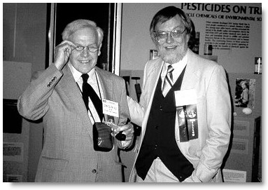
Wild and Reid in 1988 **
Picture of Dr. Wild on the top left courtesy of Dr. Wild.Adapted in part from " Stamp Vignette on Medical Science " --- John Julian Wild-Pioneer in Ultrasonography by Marc A. Shampo, Ph.D., and Robert A. Kyle, M.D. which was published in the Mayo Clinics Proceedings volume 72, page 234, 1997, and a
Press release of the Third meeting of the Federation of Ultrasound in Medicine and Biology, Brighton, England in July 1982: "the scientific discovery of sonic reflection by soft tissues and application of ultrasound to diagnostic medicine and tumour screening".
** At the History of Ultrasound Symposium in Washington DC, USA, in October 1988. Image courtesy of Dr. Eric Blackwell, reproduced with permission.
Back to History of Ultrasound in Obstetrics and Gynecology.