The use of ultrasonics in physical therapy dates back to the 1940s. The destructive ability of high intensity ultrasound had been recognised from the time of Paul Langevin when he noted destruction of school of fishes in the sea and pain induced in the hand when placed in a water tank insonated with high intensity ultrasound. In 1938, Raimar Pohlman demonstrated the 'therapeutic" effects of ultrasonic waves in human tissues. He introduced ultrasonic physiotherapy as a medical practice at the Charite in Berlin. He suggested that the power of the transducer should be limited to 5 W/cm^2, the transducer must be kept in motion, and insonifying the bone must be avoided.
In 1942, Lynn and Putnam successfully used ultrasound waves to destroy brain tissue in animals. It was considered as the earliest experiment of therapeutic ultrasound application on biological tissues. They showed that only these tissues close to the skull had been damaged and tissues in depth within the skull could be destroyed only when part of the skull was removed. William Fry and Russell Meyers performed craniotomies and used ultrasound to destroy parts of the basal ganglia in patients with Parkinsonism.
Their early experiments showed that that brain tissues deep in the skull were damaged when part of the skull was soaked in water, but the produced lesion deviated from the theoretic position. Different parts of brain tissues reacted differently to ultrasound. For example, the white matter in cat brain was susceptible to ultrasound exposure.
Peter Lindstrom reported ablation of frontal lobe tissue in moribound patients to alleviate their pain from carcinomatosis. Although some progresses had been achieved by selecting the acoustic frequencies, improving the ultrasound emission and the focusing modes, some focus deviation could not avoided and a precise abalation may not always be acheived. The acoustic intensities used could reach several hundred Watts/cm2, and the highest over ten thousand Watts/cm2. In vivo or in vitro experiments on liver, prostate and breast cancer were performed.
The 1940s saw exuberant claims made in some sectors on the effectiveness of ultrasound as an almost "cure-all" remedy, abeit the lack of much scientific evidence. This included conditons such as arthritic pains, gastric ulcers, eczema, asthma, thyrotoxicosis, haemorrhoids, urinary incontinence, elephanthiasis and even angina pectoris! Cynicism and concern over harmful tissue damaging effects of ultrasound were also mounting, which had curtailing consequences on the development of diagnostic ultrasound in the years that followed.

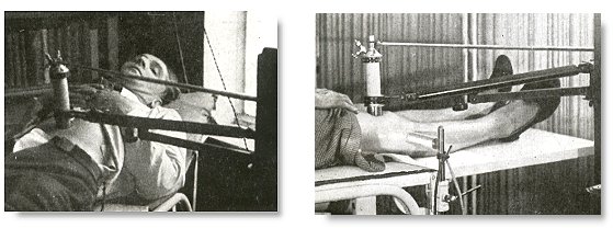
Uses of ultrasonic energy in the 1940s. Left, in gastric ulcers. Right, in arthritis*.
Thermal energy is made used of in ultrasonic therapy. When sound is propagated through tissues, the degree that any medium is exposed to heat depends on the tissue thickness. The intensity in terms of different tissue depths can be found from the equation: I = Ioe^(-alphax) where I is the intensity in tissue at depth x, Ioe is the surface intensity, and alpha is the coefficient of absorption. The frequency of the wave will also have an effect on the generation of heat. The greater the frequency, the greater the amount of energy absorbed and converted to heat according the the equation: f = 1/T where T is the period as in simple harmonic motion. Muscle and bone have been found to absorb more energy at interfaces with other heterogeneous tissues, because at these surfaces, the longitudinal waves of ultrasound are reflected and transformed into transverse waves, creating a heating effect. This happens commonly in the areas in between the muscle and bone or between the muscle and tendon. By applying ultrasonic waves to these areas, physical therapists can take advantage of this thermal affect to reduce inflammation and increase mobility in the joints.
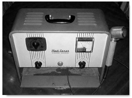
Ultrasonic therapy generator, the "Medi-Sonar" in the 1950s.
High intensity focued ultrasound (HIFU) was also applied to treat ophthalmological diseases. Lavine in 1952 reported that focusing ultrasound beams on the crystalline lens might induce the cataract. Later, some researchers destroyed the ciliary body, crystalline lens, retina, choroids using focused ultrasound and clinical experiments were conducted to treat glaucoma. Ultrasonics was also extensively used in physical and rehabilitation medicine. Jerome Gersten reported in 1953 the use of ultrasound in the treatment of patients with rheumatic arthritis. Several groups such as the Peter Wells group in Bristol, England, the Mischele Arslan group in Padua, Italy and the Douglas Gordon group in London used ultrasonic energy in the treatment of Meniere's disease.
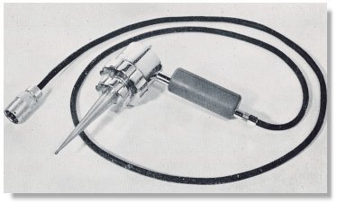
A British ultrasonic apparatus for the treatment of Meniere's disease in the late 1950s.
The use of ultrasound has gained progressive popularity in departments of Physical Medicine and Rehabilitation throughout the United States and Europe. Therapy rather than diagnosis was also the major objective of the American Institute of Ultrasound in Medicine (AIUM) in the early 1950s until the mid 1960s. The destructive nature of high intensity ultrasound also posted as an initial deterrent to it's application in medical diagnosis.
* From Andre Denier: Les Ultra-sons appliques a la medecine. 1951.
Back to History of Ultrasound in Obstetrics and Gynecology.

