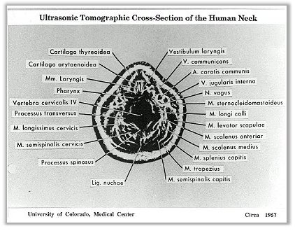
'Somagram' of the human neck obtained by Douglass Howry in 1957 using the waterbath scanner. The patient had to be sitting almost completely immersed in water up to the level of interogation and the scanning transducer moving circumferentially around the rim of the bath.
An accurate, 2D and reproducible image is produced. In the early days of pulse-echo ultrasound, the concept held was that the larger ultrasonic echoes returned by the human anatomy were those originating as specular reflections from major interfaces so that, by selectively recording these and suppressing any small echoes, it would be possible to derive outline anatomical maps or images, such as the one shown on the left. This concept was also thought to be particularly relevant in scanning the gravid uterus. The success of this approach had a strong influence on the direction of development of ultrasonic imaging in the ensuing 15 years, not least because its implementation is rather undemanding in relation to instrumental performance criteria such as signal to noise ratio and display dynamic range.In later developments, particularly after the advent of 'grey-scale' in 1975, the direction has completely shifted towards the detection (and not suppression) of small echoes in the presence of noise and subsequently to display the full dynamic range of the detected echo complex.
Source of Image: Howry D.H. (1957) Ultrasound in Biology and Medicine ed. E Kelly pp. 49-65, and from Dr. Posakony, personal communication.
Back to History of Ultrasound in Obstetrics and Gynecology.
