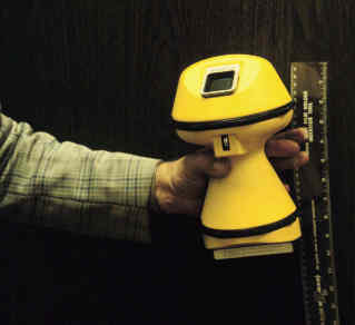
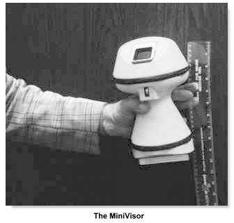
The Minivisor®, Organon Teknika (available from 1979) was a spin-off from Nicolaas Bom's laboratory. It was battery operated, shaped like a mushroom, had no wires and used a 2-inches display with an on-screen caliper system and digital readout. The transducer is fused to the bottom of the device similar to a 'large' fetal pulse detector. Juiry Wladimiroff suggested in 1980 the device would be useful for routine BPD screening. The popularity of these machines were short-lived however for several important reasons pertaining at that time: The resolution was unsatisfactory because of the available electronics. The images of the 'standard' and larger devices, as well as their overall 'portability' have seen rapid improvements round about the same time; and thirdly, realtime ultrasound has very rapidly established itself as a definitive diagnostic entity and the concern for good image information appeared to overide that of the extra portability.
The Minivisor
Schematic cross-sectional diagram indicating the major components of the Minivisor. *
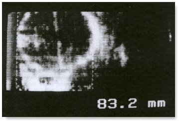
Real-time picture of the BPD showing mid-line, caliper lines superimposed on proximal and distal skull echoes. The image is 2x4 cm with a line density of 20 lines per cm. **
* from Ligtvoet C. et al. Real Time Ultrasonic Imaging with a hand-held scanner. Ultrasound in Med. & Biol., 4:91-92, 1978.
** from Wladimiroff J.W. et al. A comparative study between a linear array real-time hand-held scanner (Minivisor) and a compound scanner (Diasonograph) in the measurement of the fetal biparieal diameter. Ultrasound in Med. & Biol. 7:73-77, 1980.
Back to History of Ultrasound in Obstetrics and Gynecology.


