Martin H Wilcox was born in 1940. He graduated Bachelor of Science in Electrical Engineering (B.S.E.E.) from The Moore School of Electrical Engineering at the University of Pennsylvania, Philadelphia in 1966. In his early career he worked with physicians at the University Deparment of Radiology, testing digital computers used in radioisotope computer tomographic scanning. This introduced him to medical electronics in a hospital environment. Wilcox later joined the United TeleControl Electronics and the Penura Corporations, designing and fabricating hand-held, battery-operated doppler ultrasound diagnostic devices and automatic blood pressure measuring instruments and audiometers.
Wilcox worked at the Unirad Corporation from 1968 to 1972 and was the third employee of the company and designer of a number of their important hardware on their static B-mode scanners. Unirad, an ultrasound company started by Ray Elliott in Denver, Colorado, was a major manufacturer of ultrasonic equipment until it was sold to Technicare in 1975s. (Technicare was acquired by Johnson & Johnson in 1979). Wilcox left Unirad in '72, and started Advanced Diagnostic Research Coporation (ADR®) in Tempe, Arizona with his partner, Edward Diethrich MD. Diethrich was a well-known cardiovascular surgeon and innovator in Arizona. He established the Arizona Heart Institute in 1971. The Arizona Heart Institute Foundation was also established at around the same time to develop and support research and educational programs in cardiology. Under the leadership of Diethrich, the Institute and the Foundation have pioneered the development of preventive programs and new diagnostics and medical and surgical therapies for heart disease.
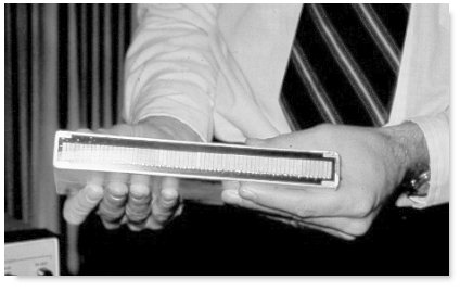
Wilcox was engineering wizard behind the invention of one of the earliest commercially available linear-array real-time scanner which first debuted in 1973. The scanner, which had set a standard for subsequent designs to follow, was the first 'good-resolution' linear-array scanner suitable for abdominal and obstetrical scanning in the commercial market. The machine's linear-array transducer contained 64 crystals in a row (3 times the number in the earlier cardiac counterparts and 3 times as long and wide), fabricated with the best material available at the time and using 'stepping' crystals techniques (see below patent claims). 
The following is part of the patent claims filed by Wilcox in 1973: 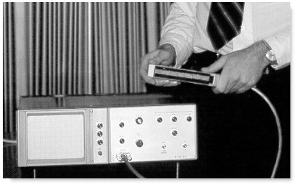
The earliest commercially available ADR scanner
The ADR probe with 64 transducer elements.
1. In a real-time ultrasonic cross-sectional imaging system, said system including
the improvement whereby the quality of said image is substantially improved in comparison to rotating transducer and fixed-array transducer systems, said improvement comprising:
- timing generator means for generating a pulsed electrical signal,
- transducer means for
- generating a pulsed ultrasonic energy beam in response to said electrical signal,
- transmitting said pulsed ultrasonic beam into a heterogenous body,
- receiving echos reflected from a moving discontinuity in said body, and
- generating an electrical echo signal in response to said received echos, and
- display means for converting said echo signal to a continuous visible image representing the moving cross-sectional structure of said heterogenous body.
N, a and B being positive integers and N 22 A, N > B.
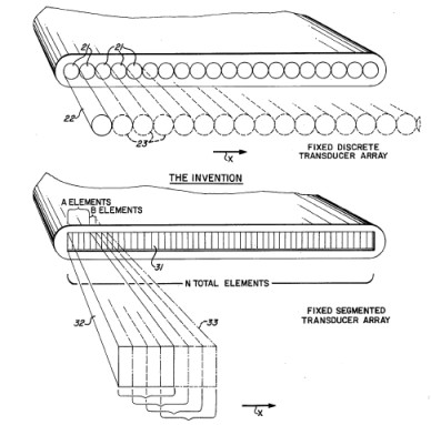
- a. said transducer means consisting of a fixed segmented elongated array formed of N discrete adjacent transverse transducer elements, said elements being large in comparison to the wave length of said pulsed ultrasonic energy beam;
- b. element counter-selector means for
- 1. transmitting identical cophased pulsed electrical signals to A selected adjacent transducer elements to generate identical cophased pulsed ultrasonic energy beams, and
- 2. sequentially pulsing selected groups of A contiguous elements as in paragraph (b) (1), each of which groups are longitudinally displaced along said elongate array, each said group being displaced B elements from the location of the immediately preceding group;
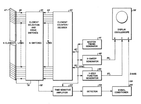
schematics of the design from Wilcox's original patent application in August 1973.
" .............. The width of each transducer segment determines the lateral resolution of the system and the number of segments determines the number of lines in the display raster. For example, in a preferred embodiment of the invention, I employ an array composed of 64 segments (N=64) each 1/8 inch wide by ?inch wide by 8 inches long. The electronic switching circuitry which controls the pulse drive and receiver selection is arranged so that four segments in the array (effectively a transducer ?inch by ?inch square) are pulsed simultaneously and allowed to receive the returning echose (A=4). After a time sufficient that all return echos have ceased, the switching circuits disconnect the first segment of the group and connect the next adjacent segment in the array (B=1), leaving the second, third and fourth segments still connected to the pulser-receiver. In effect then, the ?inch by ?inch square transducer has been moved only 1/8 inch laterally. This provides an improvement in lateral resolution four times greater than a system of discrete ?inch square transducers side by side which are sequentially pulsed. The transducer formed by the next group of 4 segments is then pulsed and the echos are received. In this manner, the entire array is scanned, producing 61 lines on the oscilloscope display tube.Depending on the application of the system, more segments with proportionally improved lateral resolution may be used. Higher frequencies permit the use of more segments, since the returning echos die out in a shorter time, allowing the next group of segments to be pulsed more rapidly. Thus, the system of the present invention retains the advantage of the discrete transducer system but adds markedly increased lateral resolution with greatly improved image quality.
The exact size of the transducer elements utilized in constructing the segmented array contemplated by my invention is not critical. Selection of the size of the transducers will depend upon the end use of the imaging system. In general, the objective in selecting the size of the transducers is to use the smallest size possible to give the best possible lateral resolution. However, as the size of the transducer is reduced, beam spreading with consequent interference becomes a problem. The relation between size of the transducer (D - width of diameter), frequency of propagation of the sound wave, and velocity of the sound wave in the medium being investigated is: Sin Q = 1.22 L/ D, where Q is the half-angle of divergence of the beam and L is the velocity of sound in the medium divided by the frequency of vibration of the sound wave. Knowing the distance from the transducer to the discontinuity, one can then calculate the beam diameter at the discontinuity. For example, the distance from the transducer to the rear wall of the heart in an average-sized patient will be approximately 15 cm and the average sound velocity in human tissue is 1538 m/sec. Using transducer elements 2.5 mm wide which operate at a frequency of 2.25 Mhz and pulsing four adjacent elements at a time (giving an effective transducer diameter of 10 mm), the half-angle of divergence will be 4.8 degrees. This divergence half-angel is close to the maximum allowable for good cardiac resolution. For comparison, in the Bom unit, the transducer diameter is about 5 mm and the half-angle is 9.4 degrees. This produces a beam diameter of 50 mm at a distance of 15 cm, leading to significant beam over-lapping and distortion and producing a relatively indistinct image........... "
Their second model the 2130 marketed in 1975 had brought the linear-array principle and the application of 'focusing techniques' to commercial fruition. The machine with 16 shades of gray-scale was a big hit in the United States and had sold over 5000 units worldwide, including Germany and other European countries. The machine was marketed in Europe under the Kranzbuhler label. In 1980 a new 3.0 MHz variable focus transducer was added on to the 2130.
The new transducer contained 506 crystal elements, boasted both mechanical and phased focusing, improved gain and reduced noise, much quieter transducer operation, and switchable focal zones. The image had twice the number of data lines and probably the best real-time resolution in the industry at that time.
" ...... Then I paid a visit to Montreal to give a lecture. Fred Winsberg, who invited me, said, 'Do you want to see the ADR real-time scanner?' It was Marty Willcox [founder of ADR] who was the one who really developed electronic real-time scanning in a commercial way. I went down and was just so bowled over with this concept of placing a linear-array transducer on the patient, seeing the fetus and its movements, and with the ease of scanning that I picked up the phone and phoned the Chief Executive of my hospital straight away and said, 'You must buy this machine, it would cost 13000 pounds.' So I ordered it straight away and took it back to London and to me it was one of the most staggering innovations in ultrasound I had experienced in my life........" - Stuart Campbell, describing his encounter with the ADR scanner in the early 1970s *.
Marty Wilcox was out of the "medical" ultrasound field from about 1982, when he and his partner Edward Diethrich sold ADR?to the Squibb Corporation? He was constrained by their sale contract from working for any medical ultrasound company for a period of five years, so Wilcox became involved with improving the state-of-the-art in underwater sonar imaging systems and eventually established a new company, Marine Sonic Technology, Ltd? based in Virginia in 1993. Much of the underwater scannner technology had nonetheless come directly from medical ultrasound. ADR?merged with ATL?(Advanced Technology laboratories? in 1984. ADR?produced the 2150 in 1980 and the last model under the ADR label, the ADR 4000 in 1982. The ADR 4000 used a 512 x 512 x 6 digital scan converter generally found in larger machines. For his inventiveness and contribution to the development of medical ultrasonography, Wilcox was awarded the Ian Donald Gold Medal for Technical Merit in 1993 by the International Society of Ultrasound in Obstetrics and Gynecology (ISUOG).
Wilcox used his experience in the medical world to design highly efficient, low power transducers which produce the extreme high quality performance underwater. In the late 1980's Wilcox designed and produced a unique underwater acoustic positioning system called SHARPS (Sonic High-Accuracy Range and Positioning System) which produces three dimensional centimeter accuracy in underwater measurements. SHARPS play a very important role in modern underwater archaeology. Marine Sonic Technology, Ltd?now makes important and highly technical underwater systems and is engaged with projects from the United States goverment, the navy and many other overseas corporations.
Pictures of Mr. Wilcox courtesy of Mr. Wilcox.Images of the early ADR scanner and meeting courtesy of Dr. Eric Blackwell, reproduced with permission.
Schematic diagrams from US Patent application, no. US1973000389958, filed August 20, 1973.
** from the the Black Sea Project.
Back to History of Ultrasound in Obstetrics and Gynecology.
